The Rat Ovaries after Erythropoietin Process
DOI:
https://doi.org/10.5530/fra.2023.2.14Keywords:
Ischemia, Erythropoietin, Ovarian epithelium edema, Ovarian congestion, Ovarian epithelium Karyorrhexis, Oophoritis, ReperfusionAbstract
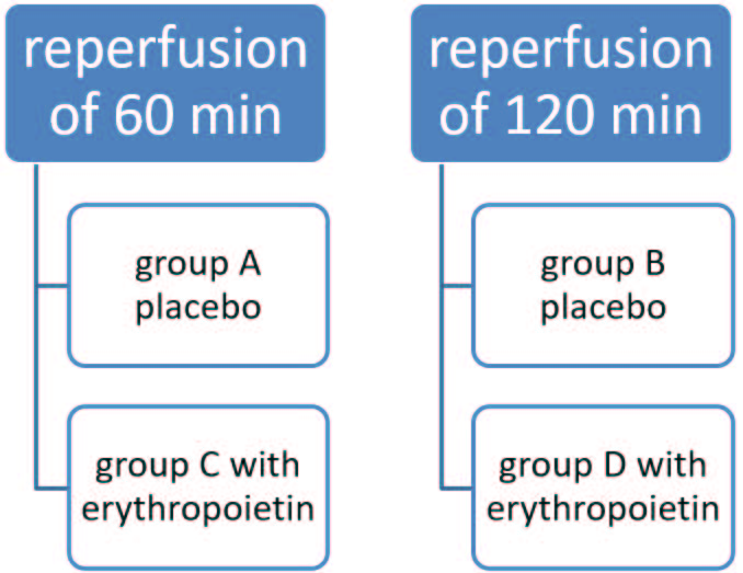
Background: This article presents an experimental model for assessment of Erythropoietin (Epo) effects produced upon post-ischemic damage of ovarian tissue. The results of study were expressed as a combined index calculated from individual pathologic scores of experimental ovarian damages. The panel included 4 distinct histologic variables, those of Ovarian Epithelium edema (OE), Ovarian Congestion (OC), Ovarian epithelium Karyorrhexis (OK) and Oophoritis (OO). Final conclusions were based on the data from 2 independent experimental series with acute ischemia-reperfusion of ovaries produced in female rats, assessing the effects of locally injected Erythropoietin (Epo). Moreover, the proinflammatory cytokine (TNFα) and Malonic Dialdehyde (MDA) were also assayed as biomarkers of oxidative stress. Materials and Methods: The study was performed in young Wistar rats. Ovarian ischemia was induced by clapping inferior aorta for 45 min after the laparotomy. Two experimental time points were chosen for assessing the OE, OC and OK, OO and TNFα, MDA scores, i.e., 60 and 120 min after starting the ovarian reperfusion. The groups A and B were served as controls, whereas the groups C and D were administered Epo intravenously. Results: The first experimental series showed that Epo has a non-significant enhancing effect for the OE and OC indexes (p-values=0.94) in a subgroup with the histologically “unchanged” state, at a grade of 0.009 [-0.258-+0.276]. The second study showed that Epo treatment was associated with a moderate, however, non-significant increase of OK and OO within the animals with “unchanged” state, grade 0.027 [-0.055-0.110] (p-values=0.50). These two studies were co-evaluated since they were obtained in the same experimental setting. Separate calculations were performed for TNFα and MDA scores showing some marginal unspecified effects of Epo. Conclusion: Epo administration was associated with a trend for enhancement of the 4 histologic variables within the “unchanged” grade group at the score of 0.018 [-0.128-+0.165] (p-value=0.80), along with some reduction of the TNFα levels by 22.89% [+15.05%] (p-value=0.12), and non-significant increase of MDA levels by 27.05% [+29.63%] (p-value=0.35). This minimal antioxidant effects should be interpreted with caution. Very likely, the antioxidant effects will be more pronounced at later observation terms.
Downloads
Metrics


